Natural history museums have entered a new stage of discovery and accessibility — one where scientists around the globe and curious folks at home can access valuable museum specimens to study, learn or just be amazed. This new era follows the completion of openVertebrate, or oVert, a five-year collaborative project among 18 institutions to create 3D reconstructions of vertebrate specimens and make them freely available online.
The team behind this endeavor, which includes scientists at the University of Washington and its Burke Museum of Natural History & Culture, published a summary of the project March 6 in the journal BioScience, offering a glimpse of how the data can be used to ask new questions and spur the development of innovative technology.
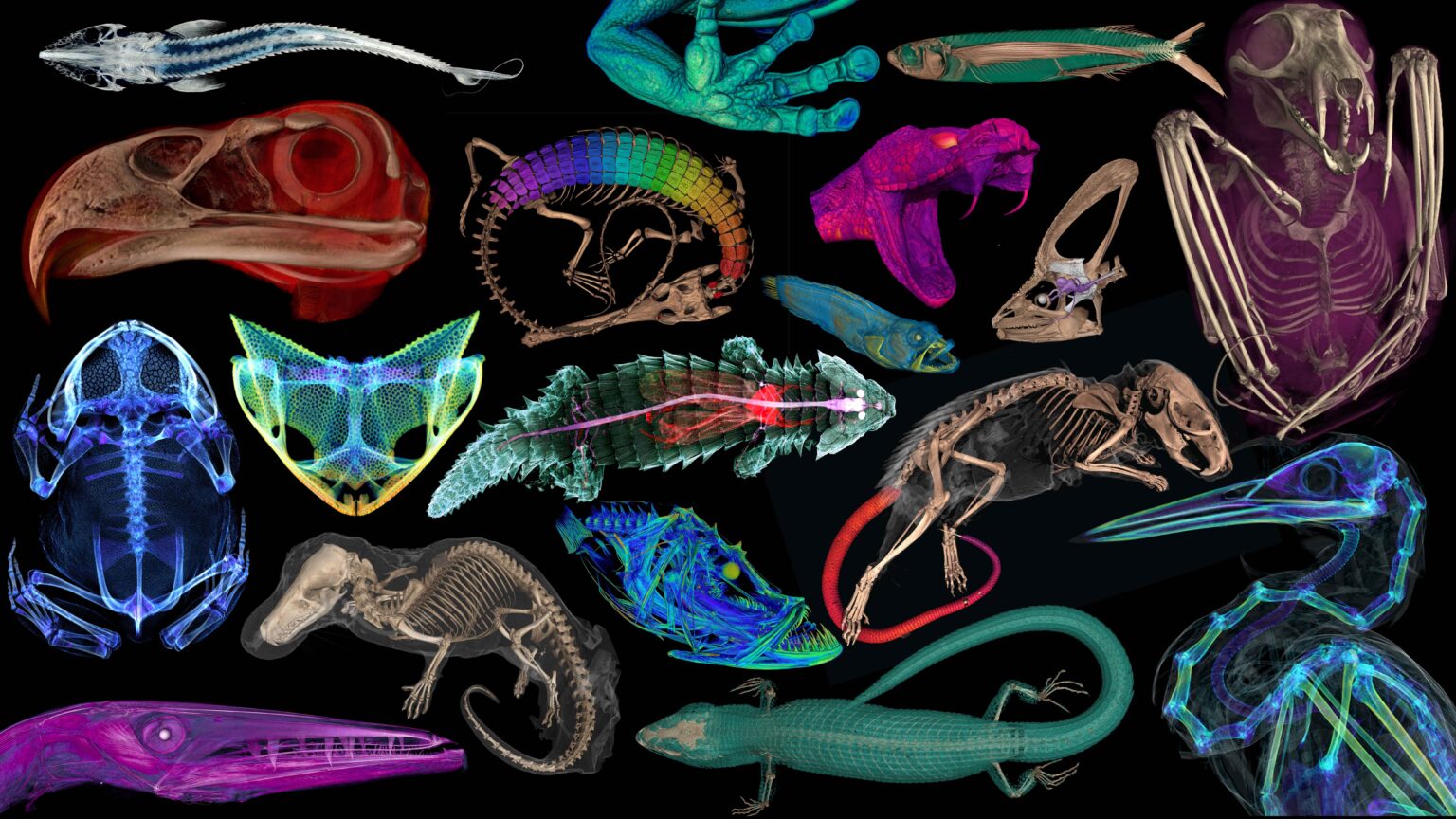
Between 2017 and 2023, oVert project members took CT scans of more than 13,000 vertebrate specimens. For the project, a team at the UW’s Friday Harbor Laboratories scanned more than 7,200 specimens — mostly fish, but also reptiles, amphibians and mammals — using the facility’s micro-CT scanner. Many of the specimens scanned at Friday Harbor came from the Burke Museum’s permanent collection. The UW team also trained more than 150 researchers, students and educators from around the world on how to CT scan specimens and analyze them for study purposes.
“It is so exciting to deposit the skeletal data for a new species in a repository where any scientist can access it,” said oVert team member Adam Summers, a UW professor of biology and of aquatic and fishery sciences, who is based at Friday Harbor Labs.
Read more at UW News »