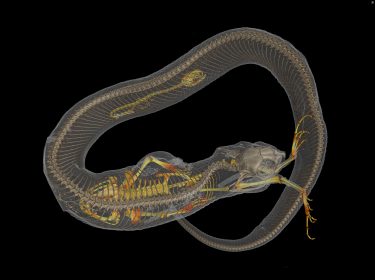
Thousands of vertebrate specimens will come off museum shelves as part of a $2.5 million project funded by the National Science Foundation. Using a CT scanner, Co-Principal Investigator Luke Tornabene, an assistant professor of aquatic and fishery sciences at UW, and his colleagues at 15 other universities will create data-rich, 3-D images of 20,000 vertebrates. All the images, part of the openVertebrate project, will be available online to researchers, educators, students and the public.
With virtual access to specimens, researchers could peel away the skin of a passenger pigeon to glimpse into its circulatory system, a class of third graders could determine a copperhead’s last meal, undergraduate students could 3-D print and compare skulls across a range of frog species and a veterinarian could plan a surgery on a giraffe in a zoo.
“In a time when museums and schools are losing natural history collections and giving up due to costs, we are recognizing the information held in these specimens is only getting more valuable,” said Tornabene, who’s also UW’s curator of fishes at the Burke Museum. “I think this project is going to help create a renaissance of the importance of natural history collections.”
https://www.facebook.com/uwnews/videos/1533270230098900/

